How PET Scans Changed Our Understanding of Alzheimer's Disease
For a long time, Alzheimer's disease was mostly diagnosed by pattern recognition. A person developed memory loss. The symptoms gradually progressed. Other causes were considered and ruled out. If the story fit, the diagnosis was often "probable Alzheimer's disease."
That was not a bad approach for its time. Clinicians still need the story. They still need to know what changed, when it changed, how it affects daily life, what medications the person takes, what the neurologic exam shows, and whether depression, sleep problems, vitamin deficiencies, thyroid disease, strokes, or other conditions might be contributing.
But Alzheimer's disease is not just a symptom pattern. It is a biological disease process in the brain. And PET imaging helped move the field from "this looks like Alzheimer's" toward "we can see evidence of the disease process while a person is still living." 1
That shift changed almost everything.
What is a PET scan?
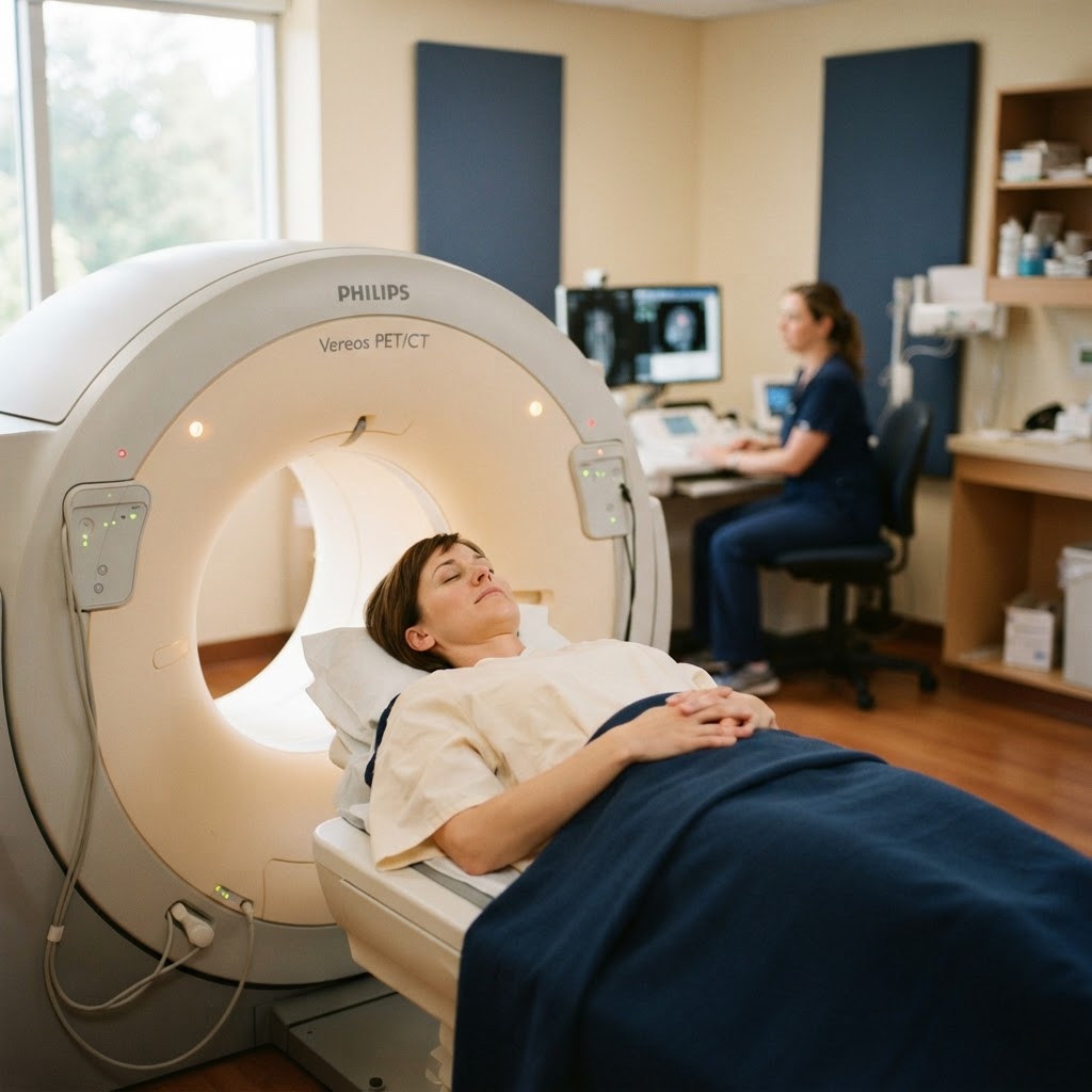
PET stands for positron emission tomography. It is a type of imaging that looks at activity or molecular changes in the body. This is different from a CT scan or MRI, which mainly show structure. CT and MRI can show things like tumors, strokes, bleeding, shrinkage, or changes in brain anatomy. PET is more about function or biology.
For a PET scan, a small amount of radioactive tracer is injected into the bloodstream. The tracer travels through the body and gives off signals that the scanner can detect. Different tracers are designed to show different things.
In dementia evaluation, the three major PET categories people usually hear about are FDG PET, amyloid PET, and tau PET. They are not interchangeable. They answer different questions.
FDG PET looks at how the brain uses glucose, which is one of the brain's main energy sources. Areas that are not functioning normally may show reduced metabolism. Amyloid PET looks for amyloid-beta plaques, one of the hallmark pathologic features of Alzheimer's disease. Tau PET looks for abnormal tau tangles, another key Alzheimer's-related pathology that tends to track more closely with symptom severity and disease progression than amyloid alone. 2,3
That sounds technical, but the simple version is this: PET scans can sometimes show evidence of Alzheimer's-related brain changes in a way that a regular MRI cannot.
Before PET, Alzheimer's was harder to confirm during life
The classic brain changes of Alzheimer's disease are amyloid plaques and tau neurofibrillary tangles. Historically, the most definitive confirmation came from examining brain tissue after death. During life, clinicians had to rely heavily on symptoms, cognitive testing, neurologic exam findings, structural imaging, and exclusion of other causes.
That created a real problem. Many conditions can mimic or overlap with Alzheimer's disease. Vascular cognitive impairment, Lewy body dementia, frontotemporal dementia, medication effects, depression, sleep apnea, and mixed pathologies can all complicate the picture. Even experienced clinicians can be wrong when they are relying only on the clinical syndrome.
PET imaging did not remove the need for clinical judgment. It did something more specific: it made it possible to detect certain Alzheimer's-related changes in the living brain. That helped researchers and clinicians understand that the biology of Alzheimer's can begin years before dementia is obvious, and that the symptoms we see in clinic are only one part of the disease story. 1
FDG PET: Looking at brain metabolism
FDG PET was one of the earlier PET tools used in dementia workups. FDG stands for fluorodeoxyglucose, a glucose-like tracer. Because brain cells use glucose for energy, FDG PET can show patterns of reduced metabolism in different brain regions.
In Alzheimer's disease, FDG PET often shows reduced metabolism in temporoparietal regions and the posterior cingulate/precuneus area. Other dementias may have different patterns. Frontotemporal dementia often affects frontal and/or anterior temporal regions. Dementia with Lewy bodies may show occipital hypometabolism and other supportive patterns. These patterns are not perfect, but they can help when the clinical picture is unclear. 4
FDG PET does not show amyloid plaques or tau tangles directly. It shows how the brain is functioning. That makes it useful in a different way. It can support a pattern of neurodegeneration and sometimes help distinguish between dementia syndromes. But it does not by itself prove that Alzheimer's pathology is present.
This distinction matters. A person can have symptoms that look like Alzheimer's disease but have a different underlying cause. A person can also have more than one process happening at the same time. FDG PET may help with pattern recognition, but amyloid and tau PET shifted the field further by letting us look more directly at the protein changes associated with Alzheimer's disease.
Amyloid PET: Seeing plaques during life
Amyloid PET was a major turning point. Instead of waiting for autopsy to confirm amyloid plaque burden, clinicians and researchers could visualize amyloid deposition in living people. In practical terms, an amyloid PET scan can help answer: Is there significant amyloid plaque in the brain?
A positive amyloid PET scan means amyloid plaques are present. That supports Alzheimer's disease biology, but it does not automatically explain every symptom. Amyloid can be present in some cognitively normal older adults, and amyloid positivity becomes more common with age. So a positive scan has to be interpreted in context.
A negative amyloid PET scan can be especially helpful. If someone has a dementia syndrome but the amyloid PET scan is negative, Alzheimer's disease is much less likely to be the primary cause of that syndrome. That can redirect the workup toward other causes, such as frontotemporal dementia, Lewy body dementia, vascular disease, psychiatric contributors, medication effects, or other neurologic conditions.
This is one of the ways PET changed dementia care. It helped separate the clinical syndrome from the underlying biology. A person may have memory loss, but memory loss does not automatically mean Alzheimer's disease. A person may have Alzheimer's pathology, but symptoms may still be shaped by other conditions in the brain or body.
Tau PET: Seeing a pattern closer to symptoms
Tau PET is newer than amyloid PET and is still more limited in routine clinical use, but it has been especially important in research. Tau tangles tend to spread through the brain in patterns that correspond more closely with cognitive symptoms and disease stage than amyloid plaque burden alone. 3
This makes intuitive sense. Amyloid is important, but amyloid can accumulate before symptoms are obvious. Tau distribution often gives a better sense of where the disease is affecting the brain and how far the process has progressed. In research settings, tau PET has helped scientists study disease staging, progression, and the relationship between brain pathology and clinical symptoms.
For patients and families, the important point is not that everyone needs tau PET. Most people do not. The point is that tau PET has helped change Alzheimer's from a diagnosis based only on symptoms to a disease that can be studied biologically across time.
PET helped redefine Alzheimer's as a biological disease
One of the biggest changes in the field is that Alzheimer's disease is increasingly defined by biology, not just by symptoms. The 2024 revised criteria from the Alzheimer's Association describe Alzheimer's disease as a biological process that can be identified by biomarkers, including amyloid PET, certain CSF tests, and accurate blood-based biomarker assays. 1
That does not mean symptoms do not matter. They absolutely do. A diagnosis that ignores the person in front of you is not good medicine. But the criteria reflect something important: Alzheimer's disease begins before dementia. The biology can be present before daily function is clearly impaired. People can be in preclinical, mild cognitive impairment, or dementia stages depending on symptoms and function, while biomarkers help identify the underlying disease process.
This is a major shift from how many people still think about Alzheimer's. Alzheimer's is often used as shorthand for dementia, but that is not quite right. Dementia describes a level of impairment. Alzheimer's disease describes a specific disease process. PET imaging helped make that distinction more visible.
PET also changed clinical trials
PET imaging transformed Alzheimer's research because it helped researchers enroll the right participants into the right studies. If a trial is testing a treatment that targets amyloid, it matters whether participants actually have amyloid pathology. Without biomarker confirmation, a study may include people whose symptoms are caused by something else. That can make it harder to know whether a treatment works.
Amyloid PET became especially important in anti-amyloid drug development. Current anti-amyloid therapies require confirmation of amyloid beta pathology before treatment is started, and amyloid PET or CSF testing are examples of ways that confirmation may be obtained. 5,6
This is not just a technical detail. It is part of why Alzheimer's research has changed so much. The field is no longer only asking, "Does this person look like they have Alzheimer's?" It is asking, "Is the biology we are targeting actually present?"
Where PET fits in a dementia workup
PET is not usually the first step in evaluating cognitive decline. A good dementia workup still begins with a careful history, collateral information from someone who knows the person well, cognitive testing, medication review, neurologic examination, screening labs, and structural imaging such as MRI or CT when appropriate.
MRI is still extremely important. It can show strokes, tumors, bleeding, hydrocephalus, patterns of atrophy, vascular disease, and other structural findings. PET does not replace that. It adds a different kind of information.
The updated appropriate use criteria for amyloid and tau PET emphasize that these scans should be used in clinical scenarios where the result is expected to help clarify diagnosis or management. They are not meant to be general screening tests for anyone who is worried about memory. They are most useful when there is cognitive impairment and uncertainty about the cause, when Alzheimer's disease is part of the differential diagnosis, or when biomarker confirmation would affect treatment decisions. 2
In plain language: PET can be very helpful, but it should answer a real clinical question.
What PET can and cannot tell you
A PET scan can provide powerful information, but it does not tell the whole story.
Amyloid PET can show whether amyloid plaque burden is present, but it cannot by itself tell you how impaired someone is or exactly how fast they will decline. Tau PET can show a pattern of tau deposition, but access is limited and interpretation still depends on the clinical context. FDG PET can show patterns of brain metabolism, but those patterns are not the same as a direct measurement of amyloid or tau.
PET also does not replace the lived history. A scan does not know whether someone is paying bills twice, getting lost, leaving the stove on, sleeping all day, acting out dreams, misreading visual information, or becoming more impulsive. Those details still matter. Sometimes they matter more than the scan.
There are also practical limitations. PET scans can be expensive. Availability varies by region. Insurance coverage can be inconsistent. CMS removed the national coverage restriction for beta-amyloid PET in 2023, allowing Medicare Administrative Contractors to make coverage decisions, but that does not mean every patient will have simple access in every setting. 7
What changed because of PET?
PET imaging changed Alzheimer's disease in several important ways.
It helped show that Alzheimer's biology can begin before dementia. It helped separate Alzheimer's disease from the broader umbrella of dementia. It helped identify people with amyloid pathology for clinical trials and newer treatments. It helped researchers understand the relationship between amyloid, tau, metabolism, symptoms, and progression. It also helped expose a truth families often learn the hard way: not all cognitive decline is the same.
That may be the most important lesson.
When someone says, "My loved one has dementia," the next question should not be only, "How bad is it?" It should also be, "What kind of dementia are we talking about, and what evidence supports that?" Sometimes the answer will be Alzheimer's disease. Sometimes it will be Lewy body dementia, frontotemporal dementia, vascular cognitive impairment, medication-related cognitive change, or a mixed picture. Often, especially in older adults, the answer is not perfectly clean.
PET scans are part of the reason we can ask better questions now.
The bigger picture
PET scans did not make dementia simple. Nothing has. But they changed what is possible. They gave researchers a way to see Alzheimer's-related biology during life. They helped move the field toward earlier and more accurate diagnosis. They helped make clinical trials more precise. And now, as blood-based biomarkers become more accurate and more widely available, PET remains one of the key tools against which many newer tests are compared. 8
For patients and families, the message is not that everyone needs a PET scan. The message is that dementia diagnosis is changing. We are moving away from vague labels and toward more specific explanations.
That is a good thing. Families deserve more than "it's dementia." They deserve to understand what that means, what it does not mean, what evidence supports the diagnosis, and what questions should come next.
References
- Jack CR Jr, Andrews JS, Beach TG, et al. Revised criteria for diagnosis and staging of Alzheimer's disease: Alzheimer's Association Workgroup. Alzheimers Dement. 2024;20(8):5143-5169. doi:10.1002/alz.13859
- Rabinovici GD, Knopman DS, Arbizu J, et al. Updated appropriate use criteria for amyloid and tau PET: a report from the Alzheimer's Association and Society for Nuclear Medicine and Molecular Imaging Workgroup. J Nucl Med. 2025;66(1). doi:10.2967/jnumed.124.268756
- Leuzy A, Palmqvist S, Mattsson-Carlgren N, et al. Considerations in the clinical use of amyloid PET and CSF biomarkers for Alzheimer's disease. Alzheimers Dement. 2025;21(2). doi:10.1002/alz.14235
- Minoshima S, Mosci K, Cross D, Thientunyakit T. 18F-FDG PET imaging in neurodegenerative dementing disorders: insights into subtype classification, emerging disease categories, and mixed dementia with copathologies. J Nucl Med. 2022;63(suppl 1):2S-12S. doi:10.2967/jnumed.121.263194
- Eisai Inc. LEQEMBI (lecanemab-irmb) injection, for intravenous use: prescribing information. Accessed June 20, 2026. https://www.leqembi.com/prescribinginformation
- Eli Lilly and Company. KISUNLA (donanemab-azbt) injection, for intravenous use: prescribing information. Accessed June 20, 2026. https://www.fda.gov/media/180803/download
- Centers for Medicare & Medicaid Services. Beta Amyloid Positron Emission Tomography in Dementia and Neurodegenerative Disease. CMS Medicare Coverage Database. Published October 13, 2023. Accessed June 20, 2026. https://www.cms.gov/medicare-coverage-database/view/ncacal-decision-memo.aspx?ncaid=308&proposed=N
- Hansson O, Blennow K, Zetterberg H, Dage J. Blood biomarkers for Alzheimer's disease in clinical practice and trials. Nat Aging. 2023;3(5):506-519. doi:10.1038/s43587-023-00403-3
This article is provided for informational and educational purposes only and does not constitute medical advice. Always consult a qualified healthcare professional for questions about diagnosis, treatment, or your personal health.